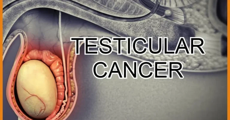
Scrotum cancer is a rare but serious condition that affects the skin of the scrotum, the pouch containing the testicles. Early detection and proper imaging play a critical role in identifying and managing this disease. This article explores the importance of scrotum cancer images in diagnosis, the various imaging modalities used, and how they aid in treatment planning.
What is Scrotum Cancer?
Scrotum cancer primarily develops as squamous cell carcinoma in the skin of the scrotum. This condition was historically one of the first occupational cancers identified and is often linked to exposure to certain chemicals and carcinogens. While not common, scrotum cancer can be aggressive if not detected and treated early.
Signs and Symptoms
Symptoms of scrotum cancer include:
- Persistent or unusual sores or lumps on the scrotum.
- Skin thickening or discoloration in the scrotal area.
- Itching, pain, or discomfort.
- Swollen lymph nodes in the groin.
Although these symptoms can be indicative of other conditions, prompt evaluation by a medical professional is essential.
The Role of Scrotum Cancer Images in Diagnosis
Imaging studies are critical tools in diagnosing scrotum cancer. These images help identify abnormalities, assess tumor extent, and plan effective treatment strategies. Below are the main imaging methods used:
1. Ultrasound Imaging
Ultrasound is the first-line diagnostic tool for scrotal abnormalities. It uses high-frequency sound waves to create detailed images of the scrotal tissues.
- Advantages:
- Non-invasive and painless.
- Provides clear differentiation between solid masses and fluid-filled cysts.
- Widely available and cost-effective.
- Limitations:
- Operator-dependent.
- Limited in visualizing surrounding lymph nodes and distant metastasis.
2. Magnetic Resonance Imaging (MRI)
MRI uses strong magnetic fields and radio waves to produce high-resolution images of the scrotum and adjacent structures. It is particularly useful for evaluating soft tissues.
- Advantages:
- Superior image clarity for soft-tissue structures.
- No exposure to ionizing radiation.
- Limitations:
- Higher cost compared to other imaging modalities.
- Longer examination time.
3. Computed Tomography (CT) Scan
CT scans are often employed for staging scrotum cancer and identifying metastasis in distant organs or lymph nodes.
- Advantages:
- Provides a comprehensive view of the body.
- Quick imaging process.
- Limitations:
- Exposure to ionizing radiation.
- Less effective for detailed soft-tissue imaging compared to MRI.
Comparing Imaging Techniques
The following table outlines the key features of each imaging modality:
| Imaging Modality | Purpose | Advantages | Drawbacks |
|---|---|---|---|
| Ultrasound | Initial assessment of scrotal masses | Cost-effective, non-invasive, no radiation | Limited for lymph node evaluation |
| MRI | Detailed imaging of soft tissues | High resolution, no radiation exposure | Expensive and less accessible |
| CT Scan | Staging and detecting metastasis | Fast, comprehensive body imaging | Involves radiation, less effective for soft tissue |
How Scrotum Cancer Images Guide Treatment
1. Early Detection
Imaging is instrumental in identifying scrotum cancer at an early stage. It helps differentiate between benign conditions and malignant tumors, ensuring timely intervention.
2. Treatment Planning
By providing detailed images of the tumor and surrounding tissues, imaging guides treatment strategies. For instance:
- Localized Tumors: Surgery or localized therapies can be planned based on tumor size and location.
- Advanced Disease: Imaging helps determine the spread of cancer, influencing the use of chemotherapy or radiation therapy.
3. Monitoring and Follow-up
Post-treatment imaging is crucial for monitoring potential recurrence and assessing treatment efficacy.
Understanding the Appearance of Scrotum Cancer on Images
Scrotum cancer often appears as irregular, thickened masses on imaging studies. Key features include:
- Ultrasound: Solid, hypoechoic (darker) lesions compared to surrounding tissue.
- MRI: Enhanced contrast highlighting tumor boundaries and adjacent involvement.
- CT Scan: Enlarged lymph nodes and potential metastasis in advanced cases.
Reducing the Risk of Scrotum Cancer
While imaging is essential for diagnosis, prevention remains a cornerstone of healthcare. Strategies to reduce risk include:
- Avoiding Carcinogens: Limiting exposure to harmful chemicals and substances associated with occupational risks.
- Maintaining Hygiene: Regular cleaning and inspection of the scrotal area.
- Prompt Medical Attention: Consulting a healthcare provider for any unusual scrotal changes.
Insights from Case Studies
Case 1: Early Detection with Ultrasound
A 45-year-old male presented with a painless scrotal lump. Ultrasound imaging revealed a solid mass, prompting early surgical intervention and successful treatment.
Case 2: MRI for Complex Cases
A patient with recurring scrotal lesions underwent an MRI, which provided detailed images of the tumor’s spread to adjacent tissues. This guided a combination of surgery and radiation therapy, improving outcomes.
Empowering Patients with Knowledge
Education is key to combating scrotum cancer. Recognizing symptoms, understanding imaging options, and seeking timely care can make a significant difference.
For more visual insights into the diagnostic process and treatment, consider exploring educational videos available online that detail the use of imaging in cancer care.
Conclusion
Scrotum cancer images play a vital role in diagnosing and managing this rare condition. From early detection with ultrasound to detailed evaluations with MRI and CT scans, imaging modalities provide critical insights into tumor characteristics and spread. By raising awareness and promoting regular medical evaluations, we can improve outcomes and reduce the impact of this disease.

